CYBERMED LIFE - ORGANIC  & NATURAL LIVING
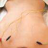
Electroacupuncture

Electroacupuncture is a form of acupuncture where a small electric current is passed between pairs of acupuncture needles. According to some acupuncturists, this practice augments the use of regular acupuncture, can restore health and well-being, and is particularly good for treating pain. There is evidence for some efficacy (when used in addition to antiemetics) in treating moderate post-chemotherapy vomiting, but not for acute vomiting or delayed nausea severity.

"Electroacupuncture is quite similar to traditional acupuncture in that the same points are stimulated during treatment. As with traditional acupuncture, needles are inserted on specific points along the body. The needles are then attached to a device that generates continuous electric pulses using small clips. These devices are used to adjust the frequency and intensity of the impulse being delivered, depending on the condition being treated. Electroacupuncture uses two needles at time so that the impulses can pass from one needle to the other. Several pairs of needles can be stimulated simultaneously, usually for no more than 30 minutes at a time."

That article adds:

"According to the principles of traditional Chinese medicine, illness is caused when chi does not flow properly throughout the body. Acupuncturists determine whether chi is weak, stagnant or otherwise out of balance, which indicates the points to be stimulated. Electroacupuncture is considered to be especially useful for conditions in which there is an accumulation of chi, such as in chronic pain syndromes, or in cases where the chi is difficult to stimulate."

Electroacupuncture is also variously termed EA, electro-acupuncture or incorporated under the generic term electrotherapy.

Electroacupuncture according to Voll (EAV) claims to measure "energy" in acupuncture points and to diagnose ailments. Some devices are registered in FDA as galvanic skin response measuring devices; they may not be used in diagnosis and treatment. Units reportedly sell for around $15,000 and are promoted for diagnosis of conditions including "parasites, food and environmental sensitivities, candida, nutritional deficiencies and much more." It is promoted for diagnosis of allergies.


  • Electroacupuncture at acupoint ST 37(Shangjuxu) improves function of the enteric nervous system in a novel mouse constipation model. 📎

    facebook Share on Facebook
    Abstract Title:

    Electroacupuncture at acupoint ST 37(Shangjuxu) improves function of the enteric nervous system in a novel mouse constipation model.

    Abstract Source:

    BMC Complement Altern Med. 2016 Oct 18 ;16(1):392. Epub 2016 Aug 18. PMID: 27756367

    Abstract Author(s):

    Chao Liang, Kaiyue Wang, Bin Xu, Zhi Yu

    Article Affiliation:

    Chao Liang

    Abstract:

    BACKGROUND:Electroacupuncture (EA) at acupoint ST 37 (Shangjuxu) has been used to alleviate gastrointestinal symptoms and improve gastrointestinal motility. However, the mechanisms by which EA affects the enteric nervous system (ENS) have scarcely been investigated. In this study, we investigated whether EA could improve ENS function.

    METHODS:A constipation model was established by gastric instillation of ice-cold saline daily for 14 days. The constipated mice were divided into two groups: the model group, which was not treated, and the EA group, which received EA at ST 37 at a frequency of 2-15 HZ and an amplitude of 1 mA for 15 min a day for 3 days. A further six mice were included as a non-constipated control group. After EA treatment, intestinal propulsion and defecation time were measured. Additionally, in jejunum, ileum and proximal colon myenteric plexus, the expressions of PGP9.5 and nNOS were measured by immunohistochemistry.

    RESULTS:The EA group demonstrated significant improvements in carbon propulsion rates and defecation time compared to model group (P < 0.05). In addition, after EA, the PGP9.5 and nNOS expression in jejunum, ileum and proximal colonic myenteric plexus was back to normal levels.

    CONCLUSION:This study suggests that EA stimulation at ST 37 is capable of ameliorating intestinal motility dysfunction, and can partly restore enteric neuron function. The ENS can participate in changes in intestinal motility by affecting inhibitory neurons.

  • Electroacupuncture at ST36 Protects ICC Networks via mSCF/Kit-ETV1 Signaling in the Stomach of Diabetic Mice. 📎

    facebook Share on Facebook
    Abstract Title:

    Electroacupuncture at ST36 Protects ICC Networks via mSCF/Kit-ETV1 Signaling in the Stomach of Diabetic Mice.

    Abstract Source:

    Evid Based Complement Alternat Med. 2017 ;2017:3980870. Epub 2017 Jan 22. PMID: 28203258

    Abstract Author(s):

    Lugao Tian, Beibei Zhu, Shi Liu

    Article Affiliation:

    Lugao Tian

    Abstract:

    Background. Electroacupuncture (EA) at ST36 has been used to regulate gastric motility and effectively improve gastric emptying in diabetic patients. Nevertheless, the specific mechanisms underlying the efficacy of this treatment remain unknown. The aim of this study was to assess the variations of interstitial cells of Cajal (ICC) and explore the changes in mSCF/KIT-ETV1 signaling in the antrum and corpus of diabetic mice after treatment with EA. Methods. Male C57BL/6 mice were randomized into five groups: control group, diabetic group (DM), diabetic-plus-sham EA group (SEA), diabetic-plus-low-frequency EA group (LEA), and diabetic-plus-high-frequency EA group (HEA). The expression levels of Ano1, c-Kit, and ETV1 were assessed by immunofluorescence in the antrum and corpus. Western blotting and PCR methods were further used to evaluate c-Kit, mSCF, and ETV1 expression. Results. (1) c-Kit and Ano1 were obviously decreased in the DM group, but c-Kit reduced much more than Ano1. (2) The mSCF, c-Kit, and ETV1 mRNA and protein levels were obviously decreased in the DM group in both the antrum and the corpus (P<0.01), but they were significantly elevated in the LEA and HEA groups (P<0.01). Conclusions. Ano1 is a reliable marker to detect ICC changes in diabetes; low- and high-frequency EA at acupoint ST36 can protect the networks of ICC possibly via normal activation of mSCF/KIT-ETV1 signaling.

  • Electroacupuncture at Zhongji (CV 3) for treatment of benign hyperplasia of prostate: a multi-central randomized controlled study

    facebook Share on Facebook
    Abstract Title:

    [Electroacupuncture at Zhongji (CV 3) for treatment of benign hyperplasia of prostate: a multi-central randomized controlled study].

    Abstract Source:

    Zhongguo Zhen Jiu. 2008 Aug;28(8):555-9. PMID: 18767576

    Abstract Author(s):

    Qing-Guo Liu, Chao-Yang Wang, Shuang Jiao, Li-Xin Tang, Ming-Hua Peng, Li-Fang Tian, Wei-Xing Ding, Xia Zhao, Shou-Kang Lu, Yong-Jun Fu, Wei-Lan Tan, Yi Qin

    Abstract:

    OBJECTIVE: To assess the role of Zhongji (CV 3) in treatment of benign hyperplasia of prostate. METHODS: Multi-central, randomized, controlled, single bland clinical method was adopted, and 276 cases were divided into an electroacupuncture (EA) group and a medication group, 138 cases in each group. The EA group were treated with EA at Zhongji (CV 3) and the medication group with oral administration of Qianliekang tablets. After treatment of 1 course, their therapeutic effects and changes of international prostate symptom (I-PSS) cumulative score, life quality index (L) cumulative score, nocturia times, urine stream state, lower abdominal symptom, maximal volume of urine flow, residual urine volume, prostatic volume, etc. Were assessed in the two groups. RESULTS: The total effective rate was 96.4% in the EA group and 86.2% in the medication group, the former being better than the latter (P<0. 01); the two groups were effective in improvement of international prostate symptom (I-PSS) cumulative score, life quality index (L) cumulative score, nocturia times, urine stream state, hypogastrium symptom, maximal volume of urine flow, residual urine volume, prostatic volume, etc. with the former better than the latter. CONCLUSION: Acupuncture at Zhongji (CV 3) has a significant therapeutic effect for treatment of benign hyperplasia of prostate.

  • Electroacupuncture at Zusanli Prevents Severe Scalds-Induced Gut Ischemia and Paralysis by Activating the Cholinergic Pathway. 📎

    facebook Share on Facebook
    Abstract Title:

    Electroacupuncture at Zusanli Prevents Severe Scalds-Induced Gut Ischemia and Paralysis by Activating the Cholinergic Pathway.

    Abstract Source:

    Evid Based Complement Alternat Med. 2015 ;2015:787393. Epub 2015 Sep 10. PMID: 26448777

    Abstract Author(s):

    Huan Wang, Lei Wang, Xian Shi, Song Qi, Sen Hu, Zhangqi Tong, Zhuhong Ma, Yan Qian, Daniela Litscher, Gerhard Litscher

    Article Affiliation:

    Huan Wang

    Abstract:

    Severe burn injuries may result in gastrointestinal paralysis, and barrier dysfunction due to gut ischemia and lowered vagus excitability. In this study we investigate whether electroacupuncture (EA) at Zusanli (ST36) could prevent severe scalds-induced gut ischemia, paralysis, and barrier dysfunction and whether the protective role of EA at ST36 is related to the vagus nerve. 35% burn area rats were divided into six groups: (a) EAN: EA nonchannel acupoints followed by scald injury; (b) EA: EA at ST36 after scald injury; (c) VGX/EA: vagotomy (VGX) before EA at ST36 and scald injury; (d) VGX/EAN: VGX before EAN and scald injury; (e) atropine/EA: applying atropine before scald injury and then EA at ST36; (f) atropine/EAN: applying atropine before scald injury and then EA at nonchannel acupoints. EA at the Zusanli point significantly promoted the intestinal impelling ratio and increased the amount of mucosal blood flow after scald injury. The plasma diamine oxidase (DAO) and intestinal permeability decreased significantly after scald injury in the EA group compared with others. However, EA after atropine injection or cervical vagotomy failed to improve intestinal motility and mucosa blood flow suggesting that the mechanism of EA may be related to the activation of the cholinergic nerve pathway.

  • Electroacupuncture Attenuates CFA-induced Inflammatory Pain by suppressing Nav1.8 through S100B, TRPV1, Opioid, and Adenosine Pathways in Mice. 📎

    facebook Share on Facebook
    Abstract Title:

    Electroacupuncture Attenuates CFA-induced Inflammatory Pain by suppressing Nav1.8 through S100B, TRPV1, Opioid, and Adenosine Pathways in Mice.

    Abstract Source:

    Sci Rep. 2017 Feb 13 ;7:42531. Epub 2017 Feb 13. PMID: 28211895

    Abstract Author(s):

    Hsien-Yin Liao, Ching-Liang Hsieh, Chun-Ping Huang, Yi-Wen Lin

    Article Affiliation:

    Hsien-Yin Liao

    Abstract:

    Pain is associated with several conditions, such as inflammation, that result from altered peripheral nerve properties. Electroacupuncture (EA) is a common Chinese clinical medical technology used for pain management. Using an inflammatory pain mouse model, we investigated the effects of EA on the regulation of neurons, microglia, and related molecules. Complete Freund's adjuvant (CFA) injections produced a significant mechanical and thermal hyperalgesia that was reversed by EA or a transient receptor potential V1 (TRPV1) gene deletion. The expression of the astrocytic marker glial fibrillary acidic protein (GFAP), the microglial marker Iba-1, S100B, receptor for advanced glycation end-products (RAGE), TRPV1, and other related molecules was dramatically increased in the dorsal root ganglion (DRG) and spinal cord dorsal horn (SCDH) of CFA-treated mice. This effect was reversed by EA and TRPV1 gene deletion. In addition, endomorphin (EM) and N(6)-cyclopentyladenosine (CPA) administration reliably reduced mechanical and thermal hyperalgesia, thereby suggesting the involvement of opioid and adenosine receptors. Furthermore, blocking of opioid and adenosine A1 receptors reversed the analgesic effects of EA. Our study illustrates the substantial therapeutic effects of EA against inflammatory pain and provides a novel and detailed mechanism underlying EA-mediated analgesia via neuronal and non-neuronal pathways.

  • Electroacupuncture Exerts Neuroprotection through Caveolin-1 Mediated Molecular Pathway in Intracerebral Hemorrhage of Rats. 📎

    facebook Share on Facebook
    Abstract Title:

    Electroacupuncture Exerts Neuroprotection through Caveolin-1 Mediated Molecular Pathway in Intracerebral Hemorrhage of Rats.

    Abstract Source:

    Neural Plast. 2016 ;2016:7308261. Epub 2016 Aug 20. PMID: 27725888

    Abstract Author(s):

    Hui-Qin Li, Yan Li, Zi-Xian Chen, Xiao-Guang Zhang, Xia-Wei Zheng, Wen-Ting Yang, Shuang Chen, Guo-Qing Zheng

    Article Affiliation:

    Hui-Qin Li

    Abstract:

    Spontaneous intracerebral hemorrhage (ICH) is one of the most devastating types of stroke. Here, we aim to demonstrate that electroacupuncture on Baihui (GV20) exerts neuroprotection for acute ICH possibly via the caveolin-1/matrix metalloproteinase/blood-brain barrier permeability pathway. The model of ICH was established by using collagenase VII. Rats were randomly divided into three groups: Sham-operation group, Sham electroacupuncture group, and electroacupuncture group. Each group was further divided into 4 subgroups according to the time points of 6 h, 1 d, 3 d, and 7 d after ICH. The methods were used including examination of neurological deficit scores according to Longa's scale, measurement of blood-brain barrier permeability through Evans Blue content, in situ immunofluorescent detection of caveolin-1 in brains, western blot analysis of caveolin-1 in brains, and in situ zymography for measuring matrix metalloproteinase-2/9 activity in brains. Compared with Sham electroacupuncture group, electroacupuncture group has resulted in a significant improvement in neurological deficit scores and in a reduction in Evans Blue content, expression of caveolin-1, and activity of matrix metalloproteinase-2/9 at 6 h, 1 d, 3 d, and 7 d after ICH (P<0.05). In conclusion, the present results suggested that electroacupuncture on GV20 can improve neurological deficit scores and reduce blood-brain barrier permeability after ICH, and the mechanism possibly targets caveolin-1/matrix metalloproteinase/blood-brain barrier permeability pathway.

  • Electroacupuncture for fatigue, sleep, and psychological distress in breast cancer patients with aromatase inhibitor-related arthralgia: a randomized trial. 📎

    facebook Share on Facebook
    Abstract Title:

    Electroacupuncture for fatigue, sleep, and psychological distress in breast cancer patients with aromatase inhibitor-related arthralgia: a randomized trial.

    Abstract Source:

    Cancer. 2014 Dec 1 ;120(23):3744-51. Epub 2014 Jul 30. PMID: 25077452

    Abstract Author(s):

    Jun J Mao, John T Farrar, Deborah Bruner, Jarcy Zee, Marjorie Bowman, Christina Seluzicki, Angela DeMichele, Sharon X Xie

    Article Affiliation:

    Jun J Mao

    Abstract:

    BACKGROUND:Although fatigue, sleep disturbance, depression, and anxiety are associated with pain in breast cancer patients, it is unknown whether acupuncture can decrease these comorbid symptoms in cancer patients with pain. The objective of this study was to evaluate the effect of electroacupuncture (EA) on fatigue, sleep, and psychological distress in breast cancer survivors who experience joint pain related to aromatase inhibitors (AIs).

    METHODS:The authors performed a randomized controlled trial of an 8-week course of EA compared with a waitlist control (WLC) group and a sham acupuncture (SA) group in postmenopausal women with breast cancer who self-reported joint pain attributable to AIs. Fatigue, sleep disturbance, anxiety, and depression were measured using the Brief Fatigue Inventory (BFI), the Pittsburgh Sleep Quality Index (PSQI), and the Hospital Anxiety and Depression Scale (HADS). The effects of EA and SA versus WLC on these outcomes were evaluated using mixed-effects models.

    RESULTS:Of the 67 randomly assigned patients, baseline pain interference was associated with fatigue (Pearson correlation coefficient [r]=0.75; P<.001), sleep disturbance (r=0.38; P=.0026), and depression (r=0.58; P<.001). Compared with the WLC condition, EA produced significant improvements in fatigue (P=.0095), anxiety (P=.044), and depression (P=.015) and a nonsignificant improvement in sleep disturbance (P=.058) during the 12-week intervention and follow-up period. In contrast, SA did not produce significant reductions in fatigue or anxiety symptoms but did produce a significant improvement in depression compared with the WLC condition (P=.0088).

    CONCLUSIONS:Compared with usual care, EA produced significant improvements in fatigue, anxiety, and depression; whereas SA improved only depression in women experiencing AI-related arthralgia.

  • Electroacupuncture for primary insomnia: a randomized controlled trial. 📎

    facebook Share on Facebook
    Abstract Title:

    Electroacupuncture for primary insomnia: a randomized controlled trial.

    Abstract Source:

    Sleep. 2009 Aug 1;32(8):1039-47. PMID: 19725255

    Abstract Author(s):

    Wing-Fai Yeung, Ka-Fai Chung, Shi-Ping Zhang, Tuan-Gee Yap, Andrew C K Law

    Abstract:

    STUDY OBJECTIVES: To evaluate the short-term efficacy and safety of electroacupuncture for the treatment of primary insomnia. DESIGN: Randomized, single-blind, placebo-controlled, parallel-group. SETTING: A university-based sleep clinic. PARTICIPANTS: Community sample of 60 Chinese adult volunteers who report having insomnia 3 or more nights per week, whose symptoms meet the DSM-IV criteria for primary insomnia for at least 3 months, and who have an Insomnia Severity Index total score of at least 15. Participants were screened with polysomnography and the Structured Clinical Interview for the DSM-IV prior to randomization. INTERVENTION: Electroacupuncture at Yintang (EX-HN3), Baihui (GV20), bilateral ear Shenmen, Sishencong (EX-HN1), and Anmian (EX) 3 times per week for 3 weeks or placebo acupuncture using Streitberger needles at the same points. MEASUREMENTS AND RESULTS: Self-reported questionnaires, 1-week sleep diaries, and 3-day actigraphy were collected at baseline and 1 week after treatment. The Insomnia Severity Index was used as the primary outcome measure. Both groups showed significant improvement compared with the pretreatment baseline. One-way analysis of covariance adjusted for baseline scores showed that there were significantly greater improvements in sleep efficiency by sleep diary and actigraphy in the electroacupuncture group. However, no significant between-group differences were observed in the Insomnia Severity Index and other outcome measures. The proportions of subjects having less than 30 minutes of wake after sleep onset and a sleep efficiency of at least 85% at the posttreatment visit were significantly higher in the electroacupuncture group. All adverse events were mild in severity. CONCLUSION: We found a slight advantage of electroacupuncture over placebo acupuncture in the short-term treatment of primary insomnia. Because of some limitations of the current study, further studies are necessary to verify the effectiveness of acupuncture for insomnia.

  • Electroacupuncture for refractory obsessive-compulsive disorder: a pilot waitlist-controlled trial.

    facebook Share on Facebook
    Abstract Title:

    Electroacupuncture for refractory obsessive-compulsive disorder: a pilot waitlist-controlled trial.

    Abstract Source:

    J Nerv Ment Dis. 2009 Aug;197(8):619-22. PMID: 19684500

    Abstract Author(s):

    Zhang-Jin Zhang, Xue-Yi Wang, Qing-Rong Tan, Gui-Xing Jin, Shao-Min Yao

    Abstract:

    A large proportion of obsessive-compulsive disorder (OCD) patients are refractory to pharmacological and cognitive-behavioral therapy. The aim of this pilot, waitlist-controlled trial was to evaluate the effectiveness of electroacupuncture (EA) as add-on therapy for treatment-resistant OCD. Nineteen patients with treatment-resistant OCD were assigned to EA treatment for 12 sessions (5 sessions per week, n = 10) or waitlist for controls (n = 9) while continuing their current anti-OCD medications. The clinical outcomes were measured using the Yale-Brown Obsessive-Compulsive Scale and the Clinical Global Impression-Severity at baseline and end point. EA additional treatment produced significantly greater improvements at end point compared with the waitlist group in reducing both Yale-Brown Obsessive-Compulsive Scale (10.2 +/- 4.2 vs. 18.8 +/- 7.4, p = 0.004) and Clinical Global Impression-Severity scores (3.0 +/- 1.1 vs. 4.4 +/- 1.1, p = 0.002). As an additional therapy, EA is effective in alleviating OCD symptoms of treatment-resistant patients. A large-scale controlled study is warranted.

  • Electroacupuncture for treatment of acute pancreatitis and its effect on the intestinal permeability of the patient

    facebook Share on Facebook
    Abstract Title:

    [Electroacupuncture for treatment of acute pancreatitis and its effect on the intestinal permeability of the patient].

    Abstract Source:

    Zhongguo Zhen Jiu. 2007 Jun;27(6):421-3. PMID: 17663105

    Abstract Author(s):

    Xin-yu Wang

    Article Affiliation:

    Section of Acupuncture&Moxibustion, General Hospital of Chinese PLA, Beijing 100853, China. This email address is being protected from spambots. You need JavaScript enabled to view it.

    Abstract:

    OBJECTIVE:To probe into effect of electroacupuncture on intestinal permeability in the patient with acute pancreatitis and the mechanism.

    METHODS:Sixty-eight cases of acute pancreatitis were randomly divided into a treatment group and a control group. The control group and the treatment group were treated with anti-infection, inhibiting secretion of pancreas, improving microcirculation and protective agent of gastric mucosa, with electroacupuncture at Zusanli (ST 36), Shangjuxu (ST 37), Gongsun (SP 4), Taichong (LR 3) and Xuanzhong (GB 39) added, twice daily for 3 days, in the treatment group. Their clinical therapeutic effects and changes of endothelin (ET), nitric oxide (NO), tumor necrosis factor (TNF-alpha) and lactulose/mannose ratio (L/M) before and after treatment were compared.

    RESULTS:The total effective rate of 86.7% in the treatment group was better than 76.3% in the control group (P<0.05). After treatment, ET, NO, TNF-alpha contents and L/M all were higher than those before treatment, with those in the treatment group being significantly lower than those in the control group (P<0.05).

    CONCLUSION:Electroacupuncture can significantly decrease permeability of intestinal mucosa in the patient with acute pancreatitis, reduce accumulation of endogenous inflammatory mediators (ET, TNF-alpha) and vascular active substance (NO) in intestinal mucosa, so as to alleviate necrosis of intestinal epithelial cells and protect the barrier of gastro-intestinal mucosa.

  • Electroacupuncture improves neurobehavioral function and brain injury in rat model of intracerebral hemorrhage.

    facebook Share on Facebook
    Abstract Title:

    Electroacupuncture improves neurobehavioral function and brain injury in rat model of intracerebral hemorrhage.

    Abstract Source:

    Brain Res Bull. 2017 Apr 7. Epub 2017 Apr 7. PMID: 28395933

    Abstract Author(s):

    Yan Zhu, Li Deng, Huajun Tang, Xiaoqing Gao, Youhua Wang, Kan Guo, Jiming Kong, Chaoxian Yang

    Article Affiliation:

    Yan Zhu

    Abstract:

    Acupuncture has been widely used as a treatment for stroke in China for a long time. Recently, studies have demonstrated that electroacupuncture (EA) can accelerate intracerebral hemorrhage (ICH)-induced angiogenesis in rats. In the present study, we investigated the effect of EA on neurobehavioral function and brain injury in ICH rats. ICH was induced by stereotactic injection of collagenase type I and heparin into the right caudate putamen. Adult ICH rats were randomly divided into the following three groups: model control group (MC), EA at non-acupoint points group (non-acupoint EA) and EA at Baihui and Dazhui acupoints group (EA). The neurobehavioral deficits of ICH rats were assessed by modified neurological severity score (mNSS) and gait analysis. The hemorrhage volume and glucose metabolism of hemorrhagic foci were detected by PET/CT. The expression levels of MBP, NSE and S100-B proteins in serum were tested by ELISA. The histopathological features were examined by haematoxylin-eosin (H&E) staining. Apoptosis-associated proteins in the perihematomal region were observed by immunohistochemistry. EA treatment significantly promoted the recovery of neurobehavioral function in ICH rats. Hemorrhage volume reduced in EA group at day 14 when compared with MC and non-acupoint EA groups. ELISA showed that the levels of MBP, NSE and S100-B in serum were all down-regulated by EA treatment. The brain tissue of ICH rat in the EA group was more intact and compact than that in the MC and non-acupoint groups. In the perihematomal regions, the expression of bcl-2 protein increased and expressions of caspase-3 and bax proteins decreased in the EA group vs MC and non-acupoint EA groups. Our data suggest that EA treatment can improve neurobehavioral function and brain injury, which were likely connected with the absorption of hematoma and regulation of apoptosis-related proteins.

  • Electroacupuncture in conscious free-moving mice reduces pain by ameliorating peripheral and central nociceptive mechanisms. 📎

    facebook Share on Facebook
    Abstract Title:

    Electroacupuncture in conscious free-moving mice reduces pain by ameliorating peripheral and central nociceptive mechanisms.

    Abstract Source:

    Sci Rep. 2016;6:34493. Epub 2016 Sep 30. PMID: 27687125

    Abstract Author(s):

    Ying Wang, Jianxun Lei, Mihir Gupta, Fei Peng, Sarah Lam, Ritu Jha, Ellis Raduenz, Al J Beitz, Kalpna Gupta

    Article Affiliation:

    Ying Wang

    Abstract:

    Integrative approaches such as electroacupuncture, devoid of drug effects are gaining prominence for treating pain. Understanding the mechanisms of electroacupuncture induced analgesia would benefit chronic pain conditions such as sickle cell disease (SCD), for which patients may require opioid analgesics throughout life. Mouse models are instructive in developing a mechanistic understanding of pain, but the anesthesia/restraint required to administer electroacupuncture may alter the underlying mechanisms. To overcome these limitations, we developed a method to perform electroacupuncture in conscious, freely moving, unrestrained mice. Using this technique we demonstrate a significant analgesic effect in transgenic mouse models of SCD and cancer as well as complete Freund's adjuvant-induced pain. We demonstrate a comprehensive antinociceptive effect on mechanical, cold and deep tissue hyperalagesia in both genders. Interestingly, individual mice showed a variable response to electroacupuncture, categorized into high-, moderate-, and non-responders. Mechanistically, electroacupuncture significantly ameliorated inflammatory and nociceptive mediators both peripherally and centrally in sickle mice correlative to the antinociceptive response. Application of sub-optimal doses of morphine in electroacupuncture-treated moderate-responders produced equivalent antinociception as obtained in high-responders. Electroacupuncture in conscious freely moving mice offers an effective approach to develop a mechanism-based understanding of analgesia devoid of the influence of anesthetics or restraints.

  • Electroacupuncture increases CB2 receptor expression on keratinocytes and infiltrating inflammatory cells in inflamed skin tissues of rats.

    facebook Share on Facebook
    Abstract Title:

    Electroacupuncture increases CB2 receptor expression on keratinocytes and infiltrating inflammatory cells in inflamed skin tissues of rats.

    Abstract Source:

    J Pain. 2010 Dec ;11(12):1250-8. Epub 2010 Jun 2. PMID: 20627823

    Abstract Author(s):

    Jing Zhang, Lin Chen, Tangfeng Su, Fuyuan Cao, Xianfang Meng, Lei Pei, Jing Shi, Hui-Lin Pan, Man Li

    Article Affiliation:

    Jing Zhang

    Abstract:

    UNLABELLED:Endogenous cannabinoids and peripheral cannabinoid CB2 receptors (CB2Rs) are involved in the antinociceptive effect of electroacupuncture (EA) on inflammatory pain. However, it remains unclear about how EA affects the expression and distribution patterns of peripheral CB2Rs in inflamed skin tissues. To study this, inflammatory pain was induced by local injection of complete Freund's adjuvant into the hindpaw of rats. The mRNA and protein levels of CB2Rs were quantified by using RTPCR and Western blotting, respectively. The distribution of CB2Rs on keratinocytes and immune cells recruited to the inflamed skin tissues was determined by using double-immunofluorescence labeling. Induction of tissue inflammation significantly increased the mRNA and protein levels of CB2Rs in the skin tissue. Also, both 2 Hz and 100 Hz EA, applied to GB30 and GB34, significantly increased the mRNA and protein levels of CB2Rs in inflamed tissues compared to the sham EA group. CB2Rimmunoreactivities were mainly distributed in keratinocytes, macrophages, and T-lymphocytes in the epidermis and dermis of the inflamed skin tissue. Inflammation caused a significant increase in the number of CB2R-immunoreactive keratinocytes, macrophages, and T-lymphocytes. Furthermore, compared to the sham EA group, EA at 2 or 100 Hz significantly increased the number of keratinocytes, macrophages, and T-lymphocytes with CB2R-immunoreactivity in the inflamed skin tissue. Therefore, our findings suggest that EA is associated with upregulation of local CB2Rs in the inflamed skin tissue. EA primarily potentiates the expression of CB2Rs on keratinocytes and infiltrating inflammatory cells at the site of inflammation.

    PERSPECTIVE:This study shows that electroacupuncture increases the CB2 receptor expression on keratinocytes and infiltrating inflammatory cells in inflammatory skin tissues. This finding provides new evidence showing the potential role of CB2 receptors in the analgesic effect of acupuncture on inflammatory pain.

  • Electroacupuncture inhibits CB1 upregulation induced by ethanol withdrawal in mice.

    facebook Share on Facebook
    Abstract Title:

    Electroacupuncture inhibits CB1 upregulation induced by ethanol withdrawal in mice.

    Abstract Source:

    Neurochem Int. 2012 Jul ;61(2):277-85. Epub 2012 May 18. PMID: 22613131

    Abstract Author(s):

    João Carlos Escosteguy-Neto, Paula Fallopa, Patrícia Varela, Renato Filev, Angela Tabosa, Jair Guilherme Santos-Junior

    Article Affiliation:

    João Carlos Escosteguy-Neto

    Abstract:

    CB1R play a role in alcohol withdrawal and in some effects of acupuncture. Interestingly, acupuncture has been used to alleviate alcohol withdrawal. Here, we investigated electroacupuncture (EA) effects during ethanol withdrawal on CB1R immunoreactivity. Male Swiss mice were daily injected with ethanol (2g/kg, i.p) (EtOH group), for 21 days. EA was performed daily during 4 days of ethanol withdrawal. The stimuli of 2 or 100 Hz were provided in two acupoints combination: Ea1 [(ST-36/Zusanli) and (PC-6/Neiguan)] or Ea2 [(DU-14/Dazhui) and (DU-20/Baihui)]. The specificity of the acupoints were assessed by the inclusion of three additional groups, Ea3 [(ST 25/Tianshu - acupoints used to other non-related disorders)], Sham1 and Sham2 (transdermic stimulation nearly to the respective acupoints). EtOH group were only handled during withdrawal and Saline group was chronically treated with Saline and handled similarly to EtOH group. One day after withdrawal the animals were perfused and their brains processed for immunohistochemistry. There was an increase of CB1R in the prefrontal cortex, striatum, hippocampus, amygdala and ventral tegmental area. The procedures used in the 2HzEa1 and 100HzEa2 groups were the most effective and specific to inhibit this CB1R upregulation. Therefore, EA inhibits CB1R upregulation seen in ethanol withdrawn mice. The specificity of acupoints stimulation depends of the encephalic nuclei, acupoints association and frequency of stimulation.

  • Electroacupuncture Inhibits NLRP3 Inflammasome Activation through CB2 Receptors in Inflammatory Pain.

    facebook Share on Facebook
    Abstract Title:

    Electroacupuncture Inhibits NLRP3 Inflammasome Activation through CB2 Receptors in Inflammatory Pain.

    Abstract Source:

    Brain Behav Immun. 2017 Aug 3. Epub 2017 Aug 3. PMID: 28782714

    Abstract Author(s):

    Fang Gao, Hong-Chun Xiang, Hong-Ping Li, Min Jia, Xiao-Li Pan, Hui-Lin Pan, Man Li

    Article Affiliation:

    Fang Gao

    Abstract:

    The therapeutic effect of electroacupuncture (EA) on inflammatory pain has been well recognized clinically. The inflammasome promotes the maturation of the inflammatory cytokines, and EA can stimulate cannabinoid CB2 receptors in inflamed tissues. In this study we investigated whether EA inhibits NLRP3 inflammasome activation through CB2 receptors and thus relieving inflammatory pain. Assay of Caspase-1 activity and western blotting revealed that complete Freund's adjuvant (CFA) injection activated the NLRP3 inflammasome in the skin tissue in rats, which was attenuated by EA treatment. Immunofluorescence labeling showed that NLRP3 inflammasome elicited by CFA in the skin macrophages were decreased by EA. Nociceptive behavioral tests demonstrated that in CB2 receptor knockout mice, the EA effects on NLRP3 inflammasomes were largely attenuated. In addition, in vitro studies in a macrophage cell line showed that CB2 receptor stimulation inhibited the NLRP3 inflammasome activation. Thus, our results suggest a novel signaling pathway through which CB2 receptors are involved in the analgesic effect of EA on inflammatory pain. Stimulation of CB2 receptors inhibits NLRP3 inflammasome activation in inflamed skin tissues. These results suggest that EA reduces the inflammatory pain by inhibiting the activation of NLRP3 inflammasome through CB2 receptors. Our findings provide novel information about the mechanisms through which EA and CB2 receptor activation reduce inflammatory pain.

  • Electroacupuncture may help motor recovery in chronic stroke survivors: a pilot study📎

    facebook Share on Facebook
    Abstract Title:

    Electroacupuncture may help motor recovery in chronic stroke survivors: a pilot study.

    Abstract Source:

    Acta Pharmacol Sin. 2005 Jun;26(6):673-8. PMID: 18712644

    Abstract Author(s):

    Wen Liu, Mukul Mukherjee, Chao Sun, Hongzeng Liu, Lisa K McPeak

    Abstract:

    Past studies have suggested that acupuncture may reduce spasticity in stroke survivors. We do not know, however, whether acupuncture may enhance the effect of strength training on motor function. This study compared upper-limb motor functional improvement in chronic stroke survivors who received a combination of acupuncture and strength training with that of subjects who received strength training alone. A total of 10 chronic stroke patients with moderate or severe wrist muscle spasticity were recruited for this study. The study used a crossover design with a random order of either combined electroacupuncture and strength training or strength training alone. Each subject received one of the two types of treatment twice a week for the first 6 weeks and switched to the other for another 6 weeks. Quantitative measurements of wrist spasticity, active wrist extension range of motion, isometric wrist strength, and clinical evaluation with Fugl-Meyer (FM) upper-limb motor scores were conducted before and after either treatment. After the combined treatment, the quantitative spasticity level, active wrist extension range of motion (increased by a mean of 16.3 degrees), and FM upper-limb motor score (increased by a mean of 4.9 points) changed significantly (p<0.01) but no significant changes were noted in isometric wrist strength. The strength training alone resulted in no significant changes to any measured variable. The results of the current study indicate that the combined acupuncture and strength training treatment reduced muscle spasticity and may have improved motor function for chronic stroke survivors with moderate or severe muscle spasticity.

  • Electroacupuncture Pretreatment Attenuates Acute Lung Injury Throughα7 Nicotinic Acetylcholine Receptor-Mediated Inhibition of HMGB1 Release in Rats after Cardiopulmonary Bypass.

    facebook Share on Facebook
    Abstract Title:

    Electroacupuncture Pretreatment Attenuates Acute Lung Injury Throughα7 Nicotinic Acetylcholine Receptor-Mediated Inhibition of HMGB1 Release in Rats after Cardiopulmonary Bypass.

    Abstract Source:

    Shock. 2017 Nov 7. Epub 2017 Nov 7. PMID: 29117064

    Abstract Author(s):

    Zhankui Wang, Lei Hou, Hao Yang, Jiaxi Ge, Shaocheng Wang, Weitian Tian, Xiangrui Wang, Zhongwei Yang

    Article Affiliation:

    Zhankui Wang

    Abstract:

    Acute lung injury is a common complication after cardiopulmonary bypass (CPB).α7 nicotinic acetylcholine receptors (α7nAChR) and α7nAChR-dependent cholinergic signaling are implicated in suppressing the release of high mobility group box 1 (HMGB1) and reducing the inflammatory response. A previous study has shown the electroacupuncture (EA) pretreatment induces tolerance against lung injury. However, the role of EA in CPB is poorly understood. This study employed EA and a rat model of CPB to determine whether EA was associated with CPB-induced lung injury. Rats were treated with EA at"Zusanli (ST36)"and"Feishu (BL13)"acupoints for 5 days before being subjected to CPB. Two hours post-CPB, samples of blood, bronchoalveolar lavage fluid (BALF) and lung tissues were processed for investigations. Our results showed that the expression ofα7nAChR in lung tissue was significantly decreased after CPB. EA pretreatment prevented the reduction in the expression of α7nAChR, EA pretreatment reduced lung edema, inhibited inflammatory cytokines release in serum and lung as well as protein concentrations in BALF and HMGB1 release following CPB, and the beneficial effects were attenuated by α-BGT. Our study demonstrates that EA pretreatment plays a protective role in CPB-induced ALI, and inhibits HMGB1 release through α7nAChR activation in rats.This is an open access article distributed under the terms of the Creative Commons Attribution-Non Commercial-No Derivatives License 4.0 (CCBY-NC-ND), where it is permissible to download and share the work provided it is properly cited. The work cannot be changed in any way or used commercially without permission from the journal. http://creativecommons.org/licenses/by-nc-nd/4.0.

  • Electroacupuncture pretreatment with different waveforms prevents brain injury in rats subjected to cecal ligation and puncture via inhibiting microglial activation, and attenuating inflammation, oxidative stress and apoptosis.

    facebook Share on Facebook
    Abstract Title:

    Electroacupuncture pretreatment with different waveforms prevents brain injury in rats subjected to cecal ligation and puncture via inhibiting microglial activation, and attenuating inflammation, oxidative stress and apoptosis.

    Abstract Source:

    Brain Res Bull. 2016 Oct 19 ;127:248-259. Epub 2016 Aug 19. PMID: 27771396

    Abstract Author(s):

    Ye Chen, Yu Lei, Li-Qun Mo, Jun Li, Mao-Hua Wang, Ji-Cheng Wei, Jun Zhou

    Article Affiliation:

    Ye Chen

    Abstract:

    Sepsis is associated with high morbidity and mortality. This study was to investigate the protective effects of electroacupuncture (EA) pretreatment with different waveforms on septic brain injury in rats and its mechanism. Male Sprague-Dawley rats were pretreated by EA with different waveforms (continuous wave, dilatational wave, or intermittent wave) at Baihui (GV20) and Tsusanli (ST36) acupoints for 30min, and underwent cecal ligation and puncture (CLP) or sham operation. The results showed that EA pretreatment with different waveforms improved survival rate, attenuated encephaledema, brain injury, neuronal apoptosis and cognitive dysfunction, and preserved blood-brain barrier (BBB). EA pretreatment decreased the production of tumor necrosis factor(TNF)-α, interleukin(IL)-6, malondialdehyde (MDA), and increased the activity of superoxide dismutase (SOD) and catalase (CAT) in serum and hippocampus at 48h after sham or CLP operation. Additionally, EA pretreatment downregulated the expressions of toll-like receptor-4 (TLR-4), nuclear factor-kappa B (NF-κB) and ionized calcium binding adaptor molecule 1(Iba 1). The effect of dilatational wave was the most significant, followed by intermittent wave, and continuous wave was relatively poor. In conclusion, our results demonstrate that EA pretreatment with three waveforms alleviates sepsis-inducedbrain injury by inhibition of microglial activation and attenuation of inflammation, oxidative stress and apoptosis. These findings suggest that EA pretreatment with dilatational wave at Baihui and Tsusanli acupoints might be a promising therapeutic strategy for relieving septic brain injury.

  • Electroacupuncture prevents endothelial dysfunction induced by ischemia-reperfusion injury via a cyclooxygenase-2-dependent mechanism: A randomized controlled crossover trial. 📎

    facebook Share on Facebook
    Abstract Title:

    Electroacupuncture prevents endothelial dysfunction induced by ischemia-reperfusion injury via a cyclooxygenase-2-dependent mechanism: A randomized controlled crossover trial.

    Abstract Source:

    PLoS One. 2017 ;12(6):e0178838. Epub 2017 Jun 7. PMID: 28591155

    Abstract Author(s):

    Seung Min Kathy Lee, Hyun Soo Kim, Jimin Park, Jong Shin Woo, Jungtae Leem, Jun Hyeong Park, Sanghoon Lee, Hyemoon Chung, Jung Myung Lee, Jin-Bae Kim, Woo-Shik Kim, Kwon Sam Kim, Weon Kim

    Article Affiliation:

    Seung Min Kathy Lee

    Abstract:

    OBJECTIVE:Exploring clinically effective methods to reduce ischemia-reperfusion (IR) injury in humans is critical. Several drugs have shown protective effects, but studies using other interventions have been rare. Electroacupuncture (EA) has induced similar protection in several animal studies but no study has investigated how the effects could be translated and reproduced in humans. This study aimed to explore the potential effect and mechanisms of EA in IR-induced endothelial dysfunction in humans.

    METHODS:This is a prospective, randomized, crossover, sham-controlled trial consisting of two protocols. Protocol 1 was a crossover study to investigate the effect of EA on IR-induced endothelial dysfunction. Twenty healthy volunteers were randomly assigned to EA or sham EA (sham). Flow mediated dilation (FMD) of the brachial artery (BA), nitroglycerin-mediated endothelial independent dilation, blood pressure before and after IR were measured. In protocol 2, seven volunteers were administered COX-2 inhibitor celecoxib (200 mg orally twice daily) for five days. After consumption, volunteers underwent FMD before and after IR identical to protocol 1.

    RESULTS:In protocol 1, baseline BA diameter, Pre-IR BA diameter and FMD were similar between the two groups (p = NS). After IR, sham group showed significantly blunted FMD (Pre-IR: 11.41± 3.10%, Post-IR: 4.49 ± 2.04%, p<0.001). However, EA protected this blunted FMD (Pre-IR: 10.96± 5.30%, Post-IR: 9.47 ± 5.23%, p = NS, p<0.05 compared with sham EA after IR). In protocol 2, this protective effect was completely abolished by pre-treatment with celecoxib (Pre-IR: 11.05± 3.27%; Post-IR: 4.20 ± 1.68%, p = 0.001).

    CONCLUSION:EA may prevent IR-induced endothelial dysfunction via a COX-2 dependent mechanism.

  • Electroacupuncture prevents ovariectomy-induced osteoporosis in rats: a randomised controlled trial. 📎

    facebook Share on Facebook
    Abstract Title:

    Electroacupuncture prevents ovariectomy-induced osteoporosis in rats: a randomised controlled trial.

    Abstract Source:

    Acupunct Med. 2012 Mar ;30(1):37-43. PMID: 22378584

    Abstract Author(s):

    Jun Zhou, Shiju Chen, Hua Guo, Lu Xia, Huifang Liu, Yuxi Qin, Chengqi He

    Article Affiliation:

    Rehabilitation Key Laboratory of Sichuan Province, Department of Rehabilitation, West China Hospital, Sichuan University, Chengdu, Sichuan 610041, People's Republic of China.

    Abstract:

    BACKGROUND:Electroacupuncture (EA) treatment has been shown to increase bone mineral density (BMD) in ovariectomised (OVX) rats; however, the underlying mechanisms remain unclear.

    OBJECTIVE:To systematically evaluate the effects of EA on OVX rats and the Wnt/β-catenin signalling pathway.

    METHODS:Three-month-old female Sprague-Dawley rats were randomly divided into three different groups (n=10 each): sham operated control (sham operated), ovariectomy (OVX) and ovariectomy with EA treatment (OVX+EA). Rats in the OVX+EA group received 12-week EA treatments.

    RESULTS:Serum bone-specific alkaline phosphatase level (p<0.01), BMD of the proximal femoral metaphysis and the fifth lumbar (L5) vertebral body (both, p<0.05) and maximum load and energy to failure of L5 vertebral body (both p<0.01) were significantly higher in the OVX+EA group than in the OVX group. Trabecular area, trabecular width and trabecular number were significantly higher in the OVX+EA group by 66.9%, 29.2% and 30.3%, respectively, than in the OVX group (all, p<0.01). Trabecular separation was 31.9% lower in the OVX+EA group than in the OVX group (p<0.01). Quantitative real-time reverse transcription polymerised chain reaction indicated that the expressions of mRNAs for low-density lipoprotein receptor-related protein 5 andβ-catenin were significantly increased in the OVX+EA group, as compared with the OVX group (p<0.01 and p<0.05, respectively).

    CONCLUSION:This study demonstrates that EA can prevent OVX-induced bone loss and deterioration of bone architecture and strength by stimulating the Wnt/β-catenin signalling pathway. These findings suggest that EA may bet a promising adjunct method for inhibiting OVX-induced osteoporosis in clinical settings.

We use cookies on our website. Some of them are essential for the operation of the site, while others help us to improve this site and the user experience (tracking cookies). You can decide for yourself whether you want to allow cookies or not. Please note that if you reject them, you may not be able to use all the functionalities of the site.