CYBERMED LIFE - ORGANIC & NATURAL LIVING
CYBERMED LIFE - ORGANIC & NATURAL LIVING
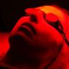 Photodynamic therapy (PDT), sometimes called photochemotherapy, is a form of phototherapy involving light and a photosensitizing chemical substance, used in conjunction with molecular oxygen to elicit cell death (phototoxicity). PDT has proven ability to kill microbial cells, including bacteria, fungi and viruses. PDT is popularly used in treating acne. It is used clinically to treat a wide range of medical conditions, including wet age-related macular degeneration, psoriasis, atherosclerosis and has shown some efficacy in anti-viral treatments, including herpes. It also treats malignant cancers including head and neck, lung, bladder and particular skin. The technology has also been tested for treatment of prostate cancer, both in a dog model and in human prostate cancer patients.
It is recognised as a treatment strategy that is both minimally invasive and minimally toxic. Other light-based and laser therapies such as laser wound healing and rejuvenation, or intense pulsed light hair removal do not require a photosensitizer. Photosensitisers have been employed to sterilise blood plasma and water in order to remove blood-borne viruses and microbes and have been considered for agricultural uses, including herbicides and insecticides.
Photodynamic therapy's advantages lessen the need for delicate surgery and lengthy recuperation and minimal formation of scar tissue and disfigurement. A side effect is the associated photosensitisation of skin tissue.

