CYBERMED LIFE - ORGANIC  & NATURAL LIVING
Magnet Therapy

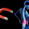
Magnet therapy, magnetic therapy, or magnotherapy is a pseudoscientific alternative medicine practice involving the use of static magnetic fields. Practitioners claim that subjecting certain parts of the body to magnetostatic fields produced by permanent magnets has beneficial health effects. These physical and biological claims are unproven and no effects on health or healing have been established. Although hemoglobin, the blood protein that carries oxygen, is weakly diamagnetic (when oxygenated) or paramagnetic (when deoxygenated) the magnets used in magnetic therapy are many orders of magnitude too weak to have any measurable effect on blood flow.

Magnet therapy is the application of the magnetic field of electromagnetic devices or permanent static magnets to the body for purported health benefits. Different effects are assigned to different orientations of the magnet.

Products include magnetic bracelets and jewelry; magnetic straps for wrists, ankles, knees, and back; shoe insoles; mattresses; magnetic blankets (blankets with magnets woven into the material); magnetic creams; magnetic supplements; plasters/patches and water that has been "magnetized". Application is usually performed by the patient.

  • A randomized, controlled trial of magnetic therapy for carpal tunnel syndrome.

    facebook Share on Facebook
    Abstract Title:

    A randomized, controlled trial of magnetic therapy for carpal tunnel syndrome.

    Abstract Source:

    Muscle Nerve. 2018 Mar 7. Epub 2018 Mar 7. PMID: 29513382

    Abstract Author(s):

    Vanessa Baute, Vahakn S Keskinyan, Erica R Sweeney, Kayla D Bowden, Allison Gordon, Janet Hutchens, Michael S Cartwright

    Article Affiliation:

    Vanessa Baute

    Abstract:

    INTRODUCTION:Magnet therapy has been proposed as a treatment for neurologic conditions. In this this trial we assessed the feasibility and efficacy of a magnet inserted into a wristband for carpal tunnel syndrome (CTS).

    METHODS:Twenty-two patients with mild to moderate CTS were randomized to wear a high-dose or low-dose"sham"magnetic wristband for 6 weeks. The primary outcome was the Symptom Severity Scale (SSS) of the Boston Carpal Tunnel Questionnaire. Secondary measures were nerve conduction studies (NCS), median nerve ultrasound, and compliance.

    RESULTS:Compliance for both groups was>90%. Improvements in the mean SSS, NCS, and median nerve ultrasound did not reach statistical significance.

    DISCUSSION:Magnet therapy via wristband is well-tolerated. Further investigations in larger populations are needed to determine efficacy. Muscle Nerve, 2018.

  • Augmenting melodic intonation therapy with non-invasive brain stimulation to treat impaired left-hemisphere function: two case studies. 📎

    facebook Share on Facebook
    Abstract Title:

    Augmenting melodic intonation therapy with non-invasive brain stimulation to treat impaired left-hemisphere function: two case studies.

    Abstract Source:

    Front Psychol. 2014 ;5:37. Epub 2014 Feb 4. PMID: 24550864

    Abstract Author(s):

    Shahd Al-Janabi, Lyndsey A Nickels, Paul F Sowman, Hana Burianová, Dawn L Merrett, William F Thompson

    Article Affiliation:

    Shahd Al-Janabi

    Abstract:

    The purpose of this study was to investigate whether or not the right hemisphere can be engaged using Melodic Intonation Therapy (MIT) and excitatory repetitive transcranial magnetic stimulation (rTMS) to improve language function in people with aphasia. The two participants in this study (GOE and AMC) have chronic non-fluent aphasia. A functional Magnetic Resonance Imaging (fMRI) task was used to localize the right Broca's homolog area in the inferior frontal gyrus for rTMS coil placement. The treatment protocol included an rTMS phase, which consisted of 3 treatment sessions that used an excitatory stimulation method known as intermittent theta burst stimulation, and a sham-rTMS phase, which consisted of 3 treatment sessions that used a sham coil. Each treatment session was followed by 40 min of MIT. A linguistic battery was administered after each session. Our findings show that one participant, GOE, improved in verbal fluency and the repetition of phrases when treated with MIT in combination with TMS. However, AMC showed no evidence of behavioral benefit from this brief treatment trial. Post-treatment neural activity changes were observed for both participants in the left Broca's area and right Broca's homolog. These case studies indicate that a combination of MIT and rTMS applied to the right Broca's homolog has the potential to improve speech and language outcomes for at least some people with post-stroke aphasia.

  • Bone healing and biochemical blood parameters after arteficial osteotomy of rabbits' femur treated by low-frequency magnetic field

    facebook Share on Facebook
    Abstract Title:

    [Bone healing and biochemical blood parameters after arteficial osteotomy of rabbits' femur treated by low-frequency magnetic field].

    Abstract Source:

    Radiat Res. 2006 May;165(5):546-52. PMID: 15503621

    Abstract Author(s):

    Z Turk

    Abstract:

    Aim of the study was to investigate the effect of magnetic fields on bone fracture healing and biochemical indices of bone turnover. Young male "White Newzealand Rabbits" were exposed after osteotomy of the femur of the hind-paw in narcosis for 31 days in a low-frequency magnetic field (field parameters: frequency 15.3 Hz, signal form: sine, magnetic field strength: 50 microT peak value). Eight animals each were assigned randomly for a single blind study to 3 groups. Controls were kept in a sham-exposition cage without magnetic field influence and were compared with a group in (Pseudo-) Helmholtz-coil arrangement with a horizontal magnetic field and a further group in a cylinder coil (vertical magnetic field). The daily duration of stay in the (sham-) exposition arrangements amounted to 30 minutes respectively. The activities of the alkaline phosphatase (AP) and creatinkinase (CK) were evaluated preoperatively, after the 19th and 31st experimental day, the callus density by means of computertomography after the 31st experimental day (two-tailed t-test). The densitometrically assessed callus densities of the magnetically treated groups were highly significant (p < 0.0001) compared with the controls. In spite of accelerated fracture healing the activities of the ALP were lower at both postoperative examinations than preoperatively, in the cylinder-coil the activity proved to be significantly lower (p < 0.05) compared with the controls after 31 days. Significant differences in CK were detected at no time. In conclusion, we confirm the positive magnetic field effects on fracture healing. The mechanisms of interaction of magnetic field and bone fracture healing remain to be clarified.

  • Combination Therapy Comprising a Static Magnetic Field with Contractility Improves Skin Wounds.

    facebook Share on Facebook
    Abstract Title:

    Combination Therapy Comprising a Static Magnetic Field with Contractility Improves Skin Wounds.

    Abstract Source:

    Tissue Eng Part A. 2018 Mar 21. Epub 2018 Mar 21. PMID: 29652610

    Abstract Author(s):

    Byeong-Wook Song, Hyunki Hong, Yu Jin Jung, Ju Hyung Lee, Bong-Soo Kim, Hoon-Bum Lee

    Article Affiliation:

    Byeong-Wook Song

    Abstract:

    Cutaneous wounds can present significant clinical problems because of abnormal healing after deep dermal damage. Despite technical advances in wound care, there are still unmet needs that result from inefficient treatment. In this study, we aimed to improve skin wound healing using a contractibility band with static magnetic field (SMF), termed a Magnetic band (Mb). To examine the effect of the Mb on wound healing, full-thickness 15 mm× 35 mm excision wounds were surgically created on the dorsum of rats. An elastic and contractile band (non-treatment), or one neodymium magnet (Nd-1) or two magnets with an elastic and contractile band (Nd-2) were topically applied to the wound daily and the wound size was measured from day 1 to 7after surgery. Nd-2 showed a significant (95%) reduction in the wound size at day 3. Histological analysis showed that pro-inflammatory cytokine levels were diminished by Nd-2, and granulation tissue and microvessels were increased compared with those in the sham group. During Mb-induced wound healing, apoptosis was significantly reduced and matrix remodeling-related factors were initially regulated. The results suggest that combination therapy comprising an SMF and an elastic and contractile band could be a promising tool to heal cutaneous wounds rapidly.

  • Complementary and Alternative Treatments for Alopecia: A Comprehensive Review📎

    facebook Share on Facebook
    Abstract Title:

    Complementary and Alternative Treatments for Alopecia: A Comprehensive Review.

    Abstract Source:

    Skin Appendage Disord. 2019 Feb ;5(2):72-89. Epub 2018 Aug 21. PMID: 30815439

    Abstract Author(s):

    Anna-Marie Hosking, Margit Juhasz, Natasha Atanaskova Mesinkovska

    Article Affiliation:

    Anna-Marie Hosking

    Abstract:

    The treatment of alopecia is limited by a lack of therapies that induce and sustain disease remission. Given the negative psychosocial impact of hair loss, patients that do not see significant hair restoration with conventional therapies often turn to complementary and alternative medicine (CAM). Although there are a variety of CAM treatment options on the market for alopecia, only a few are backed by multiple randomized controlled trials. Further, these modalities are not regulated by the Food and Drug Administration and there is a lack of standardization of bioactive in gredients in over-the-counter vitamins, herbs, and supplements. In this article, we provide a comprehensive review of the efficacy, safety, and tolerability of CAM, including natural products and mind and body practices, in the treatment of hair loss. Overall, there is a need for additional studies investigating CAM for alopecia with more robust clinical design and standardized, quantitative outcomes.

  • Effects of low frequency repetitive transcranial magnetic stimulation (rTMS) on gamma frequency oscillations and event-related potentials during processing of illusory figures in autism.

    facebook Share on Facebook
    Abstract Title:

    Effects of low frequency repetitive transcranial magnetic stimulation (rTMS) on gamma frequency oscillations and event-related potentials during processing of illusory figures in autism.

    Abstract Source:

    Lab Invest. 2008 May;88(5):529-40. Epub 2008 Mar 10. PMID: 19030976

    Abstract Author(s):

    Estate M Sokhadze, Ayman El-Baz, Joshua Baruth, Grace Mathai, Lonnie Sears, Manuel F Casanova

    Abstract:

    Previous studies by our group suggest that the neuropathology of autism is characterized by a disturbance of cortical modularity. In this model a decrease in the peripheral neuropil space of affected minicolumns provides for an inhibitory deficit and a readjustment in their signal to noise bias during information processing. In this study we proposed using low frequency transcranial magnetic stimulation (rTMS) as a way increasing the surround inhibition of minicolumns in autism. Thirteen patients (ADOS and ADI-R diagnosed) and equal number of controls participated in the study. Repetitive TMS was delivered at 0.5 Hz, 2 times per week for 3 weeks. Outcome measures based on event-related potentials (ERP), induced gamma activity, and behavioral measures showed significant post-TMS improvement. The results suggest that rTMS offers a potential therapeutic intervention for autism.

  • Effects of therapeutic magnetic stimulation on acute muscle atrophy in rats after hindlimb suspension. 📎

    facebook Share on Facebook
    Abstract Title:

    Effects of therapeutic magnetic stimulation on acute muscle atrophy in rats after hindlimb suspension.

    Abstract Source:

    Biomed Res. 2006 Feb;27(1):23-7. PMID: 16543662

    Abstract Author(s):

    Yoichi Shimada, Tsutomu Sakuraba, Toshiki Matsunaga, Akiko Misawa, Masahito Kawatani, Eiji Itoi

    Abstract:

    In most subjects with spinal cord injury, the spinal neurons below the level of injury are spared. Therefore, it is conceivable that the skeletal muscles innervated by these spinal nerves can be activated by applying therapeutic magnetic stimulation along the dorsal spine. The purpose of this study was to evaluate the ability of magnetic stimulation to prevent acute muscle atrophy in rats after hindlimb suspension. Forty adult male Wistar rats were randomly assigned to stimulated and non-stimulated (control) groups. Their hindlimbs were unweighted using a suspension method, causing muscle atrophy. In the stimulation group, magnetic stimulation (20 Hz, 60 min per day) was applied to the sciatic nerve for 10 days. After the stimulation period, the tibialis anterior (TA) and extensor digitorum longus (EDL) were surgically removed and histologically measured. The lesser diameters of type 1, 2A, and 2B muscle fibers were significantly greater in the stimulated group than in the non-stimulated group for both the TA and EDL (p < 0.05). The mean difference in lesser fiber diameter was 20% (range, 14%-27%). These results suggest that therapeutic magnetic stimulation is an effective method of preventing muscle atrophy.

  • Low intensity permanent magnets in the treatment of chronic lumbar radicular pain.

    facebook Share on Facebook
    Abstract Title:

    Low intensity permanent magnets in the treatment of chronic lumbar radicular pain.

    Abstract Source:

    J Pain Symptom Manage. 2007 Oct;34(4):434-45. Epub 2007 Jul 9. PMID: 17618081

    Abstract Author(s):

    Suzan Khoromi, Marc R Blackman, Albert Kingman, Athos Patsalides, Leigh Ann Matheny, Sandra Adams, Arthur A Pilla, Mitchell B Max

    Abstract:

    We assessed the pain-relieving efficacy of static magnetic fields produced by 200 Gauss (G) magnets compared with 50G magnets in a double-blind, randomized, two-phase crossover study in patients with chronic lumbar radicular pain. The surface field strengths of the magnets were 200 and 50G. Phase I included four random periods of two-week duration: two periods with 200G, one period with 50G, and one period of "no treatment." The magnets were positioned either vertically or horizontally in standard lumbosacral elastic corsets. Phase II consisted of two five-week periods with the most effective magnet from Phase I and its corresponding 50 or 200G device. The primary outcome was average daily leg pain score (0-10 scale) in each period of Phase II. Thirty-eight of 40 randomized patients completed Phase I, and 28 of 31 Phase II participants completed the study. In Phase I, pain scores did not differ significantly between 200 and 50G magnets. Phase II average leg pain scores tended to be lower with 200 vs. 50G magnets (3.2+/-2.1 for 200G vs. 3.9+/-2.2 for 50G magnets [P=0.08]) after excluding one unblinded patient. The relative treatment effect of the 200G magnets appeared to increase throughout the five-week period. Although these data cannot rule out a chance effect, the positive trends suggest that larger, longer-duration, sham-controlled trials with 200G magnets be considered in patients with chronic lumbar radicular pain.

  • Magnet Therapy

  • Magnet Therapy

    facebook Share on Facebook

    Magnet therapy, magnetic therapy, or magnotherapy is a pseudoscientific alternative medicine practice involving the use of static magnetic fields. Practitioners claim that subjecting certain parts of the body to magnetostatic fields produced by permanent magnets has beneficial health effects. These physical and biological claims are unproven and no effects on health or healing have been established. Although hemoglobin, the blood protein that carries oxygen, is weakly diamagnetic (when oxygenated) or paramagnetic (when deoxygenated) the magnets used in magnetic therapy are many orders of magnitude too weak to have any measurable effect on blood flow.

  • Nonpharmacological Treatments for Post-Stroke Depression: An Integrative Review of the Literature.

    facebook Share on Facebook
    Abstract Title:

    Nonpharmacological Treatments for Post-Stroke Depression: An Integrative Review of the Literature.

    Abstract Source:

    Res Gerontol Nurs. 2017 May 30:1-14. Epub 2017 May 30. PMID: 28556875

    Abstract Author(s):

    Niloufar Niakosari Hadidi, Roberta L Huna Wagner, Ruth Lindquist

    Article Affiliation:

    Niloufar Niakosari Hadidi

    Abstract:

    Stroke is the fifth leading cause of death and the number one cause of long-term disability. Seventy-five percent of annual stroke victims are older than 65. Post-stroke depression (PSD) is a common consequence of stroke, with the estimated prevalence ranging from 25% to 79%. Although several studies have investigated the impact of pharmacological interventions on PSD, there is a significant gap in knowledge regarding the efficacy of nonpharmacological measures for treatment of PSD. The purpose of the current integrative literature review was to synthesize the state of knowledge on selected nonpharmacological treatments for PSD and present findings regarding the efficacy of investigated treatments. Twenty-one studies published from 1992-2016 were identified and synthesized. Results indicated that studies demonstrating improvement in depressive symptoms included ecosystem-focused therapy, life review therapy, problem solving therapy, meridian acupressure, repetitive transcranial magnetic stimulation, music therapy, exercise, light therapy, motivational interviewing, and robotic-assisted neurorehabilitation. [Res Gerontol Nurs. 2017; x(x):xx-xx.].

  • Symptom change in women with overactive bladder after extracorporeal magnetic stimulation: a prospective trial.

    facebook Share on Facebook
    Abstract Title:

    Symptom change in women with overactive bladder after extracorporeal magnetic stimulation: a prospective trial.

    Abstract Source:

    Int Urogynecol J Pelvic Floor Dysfunct. 2007 Aug;18(8):875-80. Epub 2006 Nov 30. PMID: 17136485

    Abstract Author(s):

    Jin Ho Choe, Myung-Soo Choo, Kyu-Sung Lee

    Abstract:

    The purpose of this study was to prospectively evaluate symptom change after discontinuation of extracorporeal magnetic stimulation (EMS) in women with overactive bladder (OAB). A total of 48 women with OAB were included. We applied 10 Hz of repetitive magnetic stimulation with a "magnetic chair" for 20 min, twice weekly for 8 weeks. Changes in OAB symptoms at 2, 12, and 24 weeks after discontinuing the EMS were evaluated. Twenty-seven (56.3%) patients were cured compared with the baseline at 2 weeks: the cure rate was determined as 68.8% (33/48 patients), 56.3% (27/48), and 50% (8/16) for urgency, frequency, and urge incontinence, respectively. The mean number of voids per 24 h was decreased by 42.8% (from 14.5 +/- 4.3, to 8.3 +/- 1.5, P<0.001) at 2 weeks after treatment. Maximum voided volume did not change significantly, but the mean voided volume increased significantly after stimulation. Twenty-six (96.3%) patients among the 27 patients who achieved a cure at 2 weeks, maintained improvement at 24 weeks; the therapeutic effect on urgency, frequency, and urge incontinence persisted in 26 (78.8%) of 33 patients, 26 (96.3%) of 27 patients, and six (75%) of eight patients, respectively. There were no significant changes in urodynamic parameters. Of the 14 patients with detrusor overactivity, the condition was no longer observed in four (28.6%) patients. EMS has a beneficial effect on women with OAB. Our data suggest EMS may have a significant carry-over effect in well-selected OAB patients.

  • Transcranial magneto- and electrostimulation in patients with obesity and erectile dysfunction

    facebook Share on Facebook
    Abstract Title:

    [Transcranial magneto- and electrostimulation in patients with obesity and erectile dysfunction].

    Abstract Source:

    Vopr Kurortol Fizioter Lech Fiz Kult. 2009 Sep-Oct(5):30-3. PMID: 19886019

    Abstract Author(s):

    G N Ponomarenko, T G Bin'iash, Iu M Raĭgorodskiĭ, A S Guliaev, V A Shul'diakov, A M Kiriliuk, L Iu Vartanova

    Abstract:

    The objective of the present study was to evaluate therapeutic efficiency of transcranial magnetotherapy (TcMT) and electric stimulation (ES) included in the combined treatment of 143 patients with erectile dysfunction (ED) and abdominal obesity. The majority of the patients had waist circumference over 102 cm. An AMO-ATOS complex was used to stimulate the hypothalamic region and other brain structures. Transdermal myostimulation of the abdominal and femoral regions was achieved with a Miovolna device. It was shown that both TcM and ES improved lipid metabolism and erectile function; moreover, they exerted hypotensive and sedative action. Specifically, the testosterone level in the patients increased by a mean of 27% compared with the pre-treatment values while the number of patients complaining of erectile dysfunction decreased by 31%.

We use cookies on our website. Some of them are essential for the operation of the site, while others help us to improve this site and the user experience (tracking cookies). You can decide for yourself whether you want to allow cookies or not. Please note that if you reject them, you may not be able to use all the functionalities of the site.