CYBERMED LIFE - ORGANIC  & NATURAL LIVING

Chelation Therapy EDTA

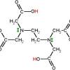
Chelation therapy is a medical procedure that involves the administration of chelating agents to remove heavy metals from the body. Chelation therapy has a long history of use in clinical toxicology and remains in use for some very specific medical treatments, although it is administered under very careful medical supervision due to various inherent risks.

Chelation therapy must be administered with care as it has a number of possible side effects, including death. In response to increasing use of chelation therapy as alternative medicine and in circumstances in which the therapy should not be used in conventional medicine, various health organizations have confirmed that medical evidence does not support the effectiveness of chelation therapy for any purpose other than the treatment of heavy metal poisoning. Over-the-counter chelation products are not approved for sale in the United States.

  • Efficacy of Chelation Therapy to Remove Aluminium Intoxication

    facebook Share on Facebook
    Abstract Title:

    Efficacy of Chelation Therapy to Remove Aluminium Intoxication

    Abstract Author(s):
    Alessandro FulgenziRachele De GiuseppeFabrizia BamontiDaniele ViettiMaria Elena Ferrero
     
    Abstract:

    There is a distinct correlation between aluminium (Al) intoxication and neurodegenerative diseases (ND). We demonstrated how patients affected by ND showing Al intoxication benefit from short-term treatment with calcium disodium ethylene diamine tetraacetic acid (EDTA) (chelation therapy). Such therapy further improved through daily treatment with the antioxidant Cellfood. In the present study we examined the efficacy of long-term treatment, using both EDTA and Cellfood. Slow intravenous treatment with the chelating agent EDTA (2 g/10 mL diluted in 500 mL physiological saline administered in 2 h) (chelation test) removed Al, which was detected (using inductively coupled plasma mass spectrometry) in urine samples collected from patients over 12 h. Patients that revealed Al intoxication (expressed in μg per g creatinine) underwent EDTA chelation therapy once a week for ten weeks, then once every two weeks for a further six or twelve months. At the end of treatment (a total of 22 or 34 chelation therapies, respectively), associated with daily assumption of Cellfood, Al levels in the urine samples were analysed. In addition, the following blood parameters were determined: homocysteine, vitamin B12, and folate, as well as the oxidative status e.g. reactive oxygen species (ROS), total antioxidant capacity (TAC), oxidized LDL (oxLDL), and glutathione. Our results showed that Al intoxication reduced significantly following EDTA and Cellfood treatment, and clinical symptoms improved. After treatment, ROS, oxLDL, and homocysteine decreased significantly, whereas vitamin B12, folate and TAC improved significantly. In conclusion, our data show the efficacy of chelation therapy associated with Cellfood in subjects affected by Al intoxication who have developed ND.

  • Chelation Therapy EDTA

  • EDTA chelation therapy, without added vitamin C, decreases oxidative DNA damage and lipid peroxidation📎

    facebook Share on Facebook
    Abstract Title:

    EDTA chelation therapy, without added vitamin C, decreases oxidative DNA damage and lipid peroxidation.

    Abstract Source:

    Altern Med Rev. 2009 Mar;14(1):56-61. PMID: 19364193

    Abstract Author(s):

    Anne Marie Roussel, Isabelle Hininger-Favier, Robert S Waters, Mireille Osman, Karen Fernholz, Richard A Anderson

    Abstract:

    Chelation therapy is thought to not only remove contaminating metals but also to decrease free radical production. However, in standard ethylene diamine tetracetic acid (EDTA) chelation therapy, high doses of vitamin C with potential pro-oxidant effects are often added to the chelation solution. The authors demonstrated previously that the intravenous administration of the standard chelation cocktail, containing high amounts of vitamin C, resulted in an acute transitory pro-oxidant burst that should be avoided in the treatment of pathologies at risk of increased oxidative stress such as diabetes and cardiovascular disease. The current study was designed to determine the acute and chronic biochemical effects of chelation therapy on accepted clinical, antioxidant variables. An EDTA chelation cocktail not containing ascorbic acid was administered to six adult patients for five weeks (10 sessions of chelation therapy); antioxidant indicators were monitored. Immediately after the initial chelation session, in contrast with the data previously reported with the standard cocktail containing high doses of vitamin C, none of the oxidative stress markers were adversely modified. After five weeks, plasma peroxide levels, monitored by malondialdehyde, decreased by 20 percent, and DNA damage, monitored by formamidopyrimidine-DNA glycosylase (Fpg) sensitive sites, decreased by 22 percent. Remaining antioxidant-related variables did not change. In summary, this study demonstrates that multiple sessions of EDTA chelation therapy in combination with vitamins and minerals, but without added ascorbic acid, decreases oxidative stress. These results should be beneficial in the treatment of diseases associated with increased oxidative stress such as diabetes and cardiovascular diseases.

  • Effect of glutathione on the cadmium chelation of EDTA in a patient with cadmium intoxication.

    facebook Share on Facebook
    Abstract Title:

    Effect of glutathione on the cadmium chelation of EDTA in a patient with cadmium intoxication.

    Abstract Source:

    Hum Exp Toxicol. 2010 Apr 22. Epub 2010 Apr 22. PMID: 20413561

    Abstract Author(s):

    Hyo-wook Gil, Eun-jung Kang, Kwon-hyun Lee, Jong-oh Yang, Eun-young Lee, Sae-yong Hong

    Article Affiliation:

    Department of Internal Medicine, Soonchunhyang University Cheonan Hospital, Cheonan City, South Korea.

    Abstract:

    In order to evaluate the efficiency and renal protective effects of glutathione during Ca(++)-EDTA chelation therapy for chronic cadmium intoxication, we measured the renal excretion of cadmium, beta2-microglobulin, proteinuria, and hematuria during intravenous administration of glutathione with Ca(++)-EDTA in a 54-year-old patient with chronic cadmium intoxication. We administered 500 mg of Ca(++)-EDTA and 50 mg/kg of glutathione alone or in 1 L of normal saline over the next 24 hours and repeated this over 12 consecutive days. During the first 3 days, the basal levels (only saline administration) were determined; during the second 3 days, Ca(++)-EDTA only was administered, for the third sequence of 3 days, Ca(++)-EDTA with glutathione was provided, and for the last 3 days, glutathione alone was given. One month later, the same protocol was repeated. There were six blood and urine samples to analyze in each group. The blood cadmium level was higher when the EDTA was infused together with glutathione (7.44 +/- 0.73 mug/L, p<0.01) compared to the basal level of 4.6 +/- 0.44 mug/L. Also, the renal cadmium excretion was significantly higher in the EDTA with glutathione group than in the basal group (23.4 +/- 15.81 mug/g creatinine vs 89.23 +/- 58.52 mug/g creatinine, p<0.01). There was no difference in the protein/creatinine and beta2-microglobulin/creatinine ratio in the urine (p>0.05) among the groups. Furthermore, microhematuria and proteinuria did not develop over the observation period of 6 months. These results suggest that glutathione administration with EDTA might be an effective treatment modality for patients with cadmium intoxication.

  • Effectiveness of chelation therapy with time after acute uranium intoxication.

    facebook Share on Facebook
    Abstract Title:

    Effectiveness of chelation therapy with time after acute uranium intoxication.

    Abstract Source:

    Fundam Appl Toxicol. 1990 Jan;14(1):88-95. PMID: 2307325

    Abstract Author(s):

    [No authors listed]

    Abstract:

    The effect of increasing the time interval between acute uranium exposure and chelation therapy was studied in male Swiss mice. Gallic acid, 4,5-dihydroxy-1,3- benzenedisulfonic acid (Tiron), diethylenetriaminepentaacetic acid (DTPA), and 5-aminosalicylic acid (5-AS) were administered ip at 0, 0.25, 1, 4, and 24 hr after sc injection of 10 mg/kg of uranyl acetate dihydrate. Chelating agents were given at doses equal to one-fourth of their respective LD50 values. Daily elimination of uranium into urine and feces was determined for 4 days after which time the mice were killed, and the concentration of uranium was measured in kidney, spleen, and bone. The excretion of uranium was especially rapid in the first 24 hr. Treatment with Tiron or gallic acid at 0, 0.25, or 1 hr after uranium exposure significantly increased the total excretion of the metal. In kidney and bone, only administration of Tiron at 0, 0.25, or 1 hr after uranium injection, or gallic acid at 1 hr after uranium exposure significantly reduced tissue uranium concentrations. Treatment at later times (4 to 24 hr) did not increase the total excretion of the metal and did not decrease the tissue uranium concentrations 4 days after uranyl acetate administration. The results show that the length of time before initiating chelation therapy for acute uranium intoxication greatly influences the effectiveness of this therapy.

  • Effectiveness of treatment of calcific tendinitis of the shoulder by disodium EDTA📎

    facebook Share on Facebook
    Abstract Title:

    Effectiveness of treatment of calcific tendinitis of the shoulder by disodium EDTA.

    Abstract Source:

    Arthritis Rheum. 2009 Jan 15;61(1):84-91. PMID: 19116968

    Abstract Author(s):

    Angelo Cacchio, Elisabetta De Blasis, Piergiorgio Desiati, Giorgio Spacca, Valter Santilli, Fosco De Paulis

    Article Affiliation:

    San Salvatore Hospital of L'Aquila, Via L. Natali 1, 67100 L'Aquila, Italy. This email address is being protected from spambots. You need JavaScript enabled to view it.

    Abstract:

    OBJECTIVE: To evaluate the effectiveness of disodium EDTA administration in the treatment of calcific tendinitis of the shoulder.

    METHODS: Eighty patients with radiographically verified calcific tendinitis of the shoulder were enrolled between September 2001 and October 2003. Patients were randomly assigned to either a study group (n = 40) or a control group (n = 40). Pain and functional level were evaluated before and after treatment and at 1-year followup. Radiographic modifications in calcifications were evaluated before and after treatment. Disodium EDTA was administered through single needle mesotherapy and 15 minutes of pulsed-mode 1 MHz-ultrasound.

    RESULTS: The study group displayed improvement in all of the parameters analyzed after treatment and at the 1-year followup. Calcifications disappeared completely in 62.5% of the patients in the study group and partially in 22.5%; calcifications partially disappeared in only 15% of the patients in the control group, and none displayed a complete disappearance.

    CONCLUSION: Our results suggest that the use of disodium EDTA for the management of calcific tendinitis of the shoulder is safe and effective, leading to a significant reduction in pain, improvement in shoulder function, and disappearance of calcifications after 4 weeks, without adverse effects.

  • Should EDTA chelation therapy be used instead of long-term clopidogrel plus aspirin to treat patients at risk from drug-eluting stents? ?

    facebook Share on Facebook
    Abstract Title:

    Should EDTA chelation therapy be used instead of long-term clopidogrel plus aspirin to treat patients at risk from drug-eluting stents?

    Abstract Source:

    Altern Med Rev. 2007 Jun;12(2):152-8. PMID: 17604460

    Abstract Author(s):

    L Terry Chappell

    Abstract:

    The recently discovered increased risk of blood clots, leading to myocardial infarction and sudden death beginning six months after medicated stents are implanted in patients following percutaneous transluminal coronary angioplasty (PTCA), has left cardiologists pondering what course of action to take. The purpose of adding implanted medication to a stent is to prevent thrombin accumulation and restenosis. However, these stents may increase, rather than decrease, the risk. Although long-term treatment with clopidogrel bisulfate (Plavix) plus aspirin for at least 12 months has been suggested as a preventive treatment, there is no evidence from randomized, controlled trials that this treatment is effective for more than six months. Clopidogrel also increases the risk of major bleeding episodes. The author served as the primary investigator for a study that showed cardiovascular patients treated with EDTA chelation therapy had a lower rate of subsequent cardiac events, including myocardial infarction and death, than those treated with cardiac medications, PTCA, or coronary artery bypass graft (CABG). The data also indicated chelation therapy might be effective in preventing thrombosis and cardiac events from stent implantation. There is evidence EDTA chelation therapy might prevent hypercoagulability resulting from the placement of stents, although not specifically medicated stents. Based on the limited data currently available, intravenous EDTA may be safe and effective for treating patients who have implanted medicated stents. Prospective clinical trials are needed, and EDTA should be included in those trials.

  • The usefulness of chelation therapy for the remission of symptoms caused by previous treatment with mercury-containing pharmaceuticals: a case report📎

    facebook Share on Facebook
    Abstract Title:

    The usefulness of chelation therapy for the remission of symptoms caused by previous treatment with mercury-containing pharmaceuticals: a case report.

    Abstract Source:

    Cases J. 2009 Nov 18;2:199. PMID: 19946446

    Abstract Author(s):

    Serafina Corsello, Alessandro Fulgenzi, Daniele Vietti, Maria Elena Ferrero

    Abstract:

    INTRODUCTION: A great deal of data regarding the toxicology of mercury has been recently reported. Although the most common human exposures to mercury are currently mercury vapour from amalgam tooth fillings, methylmercury from seafood and ethylmercury as a preservative in vaccines, in the past mercury compounds have been used in the treatment of syphilis.

    CASE PRESENTATION: Mercury intoxication was found in a 67 year-old Italian man affected by neurological symptoms of apparently unknown origin. The patient developed syphilis forty years ago and then underwent therapy with mercurials to treat his chronic bacterial infection. We treated the patient with disodium edetate chelation therapy. Six months after the beginning of the therapy, the patient's neurological symptoms began to decrease, and were completely cured after two years of therapy.

    CONCLUSION: This case supports the use of chelation therapy with disodium edetate to remove damages caused by mercury intoxication.

We use cookies on our website. Some of them are essential for the operation of the site, while others help us to improve this site and the user experience (tracking cookies). You can decide for yourself whether you want to allow cookies or not. Please note that if you reject them, you may not be able to use all the functionalities of the site.